New Brain Aging Study Questions the Role of Common Omega-3
Scientists find that EPA (eicosapentaenoic acid), an omega-3 fatty acid found in fish oil, impairs brain healing after injury in mice.
Highlights:
- Fish oil contributes to memory loss in mice with traumatic brain injury.
- Fish oil also contributes to neurodegenerative features in mice with traumatic brain injury.
- In the brain tissue of older adults with brain injury, there is an abnormal accumulation of fats, suggesting the abnormalities found in mice could translate to humans.
Traumatic brain injury (TBI), usually caused by external blunt force, disrupts normal brain function. It can gradually progress over decades, increasing the risk of dementia. Older adults have the highest rates of TBI-related hospitalizations and deaths. It follows that preventing TBI in older adults, which usually occurs from falls, can potentially increase the chances of a healthier and longer life.
There are currently no effective treatments for TBI, highlighting the importance of preventing or slowing its progression. In a new study, published in Cell Reports, researchers from the Medical University of South Carolina have found that fish oil may do the opposite. They show that EPA, a fat found in fish oil, impairs repair following brain injury. These findings suggest that EPA may be harmful to individuals with TBI.
Fish Oil and TBI Contribute to Memory Loss
How TBI progresses into cognitive decline and dementia remains unclear. To see how fish oil may be involved, the South Carolina researchers fed mice a high-fat diet (HFD) containing fish oil (FO). The HFD included EPA and DHA (docosahexaenoic acid), which are the essential omega-3 fatty acids found in FO. After one month of the FO-supplemented HFD (FO-Supp HFD), the researchers induced TBI.
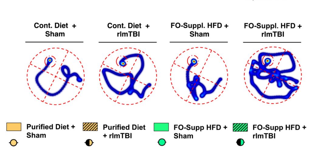
The researchers next assessed the mice’s memory using a common test known as the Morris water maze. Mice were trained to find the location of an escape platform in a pool of water. After training, the platform was removed for the testing session. Mice without TBI on a normal diet, as well as mice with TBI on a normal diet, and mice without TBI on a FO-Supp HFD, remembered and found the location of the platform relatively quickly. However, the TBI mice on the FO-Supp HFD relied on searching the perimeter of the maze before finally finding the correct location. These findings suggest that the FO-Supp HFD contributes to memory loss.
Fish Oil and TBI Contribute to Neurodegeneration
The blood vessels of the brain deliver oxygen and nutrients to neurons and other brain cells while clearing waste. The brain’s blood vessels are particularly susceptible to injury following TBI. To examine the effects of FO on the brain’s vasculature, the researchers examined the brain tissue of TBI mice fed the FO-Supp HFD. They found that the FO-Supp HFD decreased neuron size and increased aggregates of proteins associated with Alzheimer’s disease called tau.
The FO-Supp HFD also led to abnormalities in brain vasculature, including blood vessel narrowing. Moreover, the FO-Supp HFD significantly reduced blood flow in response to stimulating the mice’s whiskers. The changes in neuronal size and alterations in blood flow were not accompanied by changes in neuronal connections or blood vessel leakiness. These findings suggest that FO contributes to subclinical neurodegeneration, which may explain the cognitive decline following TBI.
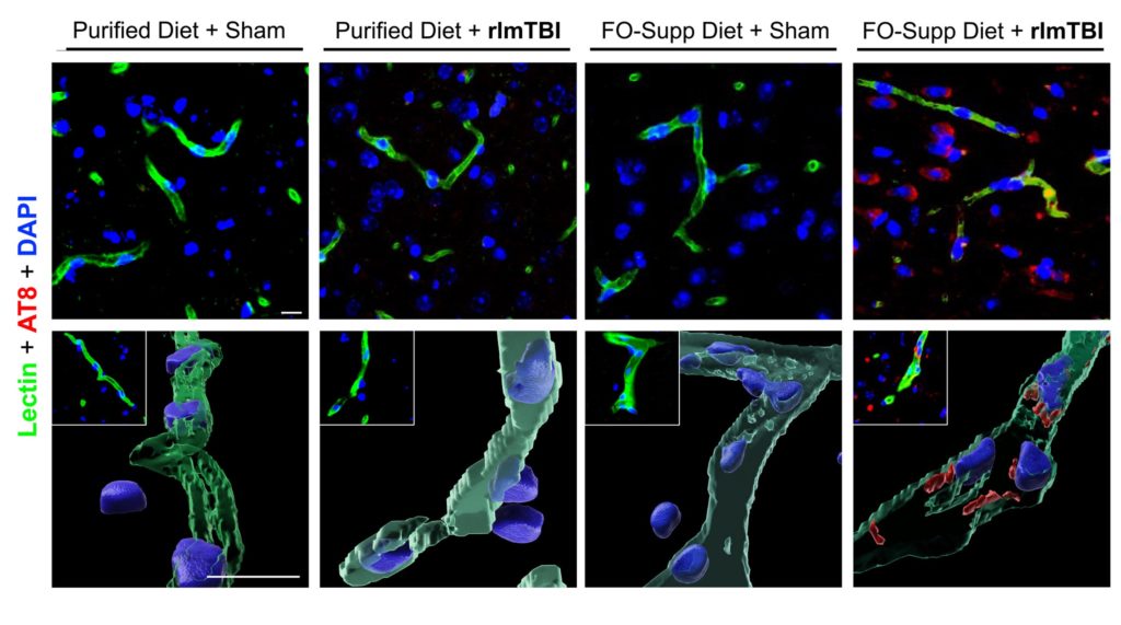
Further experimentation revealed that the EPA, but not DHA in the fish oil, promoted vasculature disruptions. EPA was shown to reduce the formation of new blood vessels and destabilize the communication between the cells that line blood vessel walls. The EPA also disrupted the wound-healing process. The findings suggest that, with TBI, EPA but not DHA hinders the repair process, potentially contributing to neurodegeneration and cognitive decline.
Abnormal Fatty Acid Buildup Found in People with Brain Injury
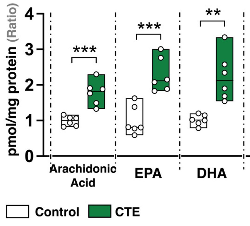
CTE (chronic traumatic encephalopathy) is a neurodegenerative disease caused by repetitive head trauma. To determine the relevance of their findings in humans, the South Carolina researchers examined brain tissue from CTE patients above the age of 75. They found a 150% increase in EPA and DHA within the brain tissue of CTE patients, which was compared to the brain tissue of healthy older adults. CTE patients also showed tau protein buildup and signs of impaired brain-blood vessel architecture, like in the mice.
Avoiding EPA
The study suggests that EPA contributes to maladaptive brain restructuring following TBI. Both EPA and DHA are incorporated into cell membranes, which allows cells to function optimally. The South Carolina researchers found signs of abnormal fat metabolism in the brain tissue of TBI mice and CTE patients, suggesting that EPA may be aberrantly incorporated into cells post-injury. However, more studies are needed to clarify how EPA may lead to cognitive decline and neurodegeneration following TBI. Until then, it may be beneficial for those with TBI to avoid EPA. Individuals without TBI are advised not to avoid EPA or DHA, as these omega-3 fatty acids are associated with a lower risk of neurodegeneration, cognitive decline, and dementia.
Model: C57BL/6J mice
Dosage: A cyclic high-fat diet containing fish oil (26.3 g/kg EPA and 20.5 g/kg DHA) every third day for 6 months
 Comments