NMN Protects Against Corneal Cell Death in Damaged Eyes
Losing nerve supplies in our window to the world — cornea — may cloud our sights and even cut out the light. But scientists found that NAD+ replenishment may be a possible intervention.
Our window to the world is the cornea, a transparent dome-like layer of cells that sits directly on top of our iris and pupil, allowing light to enter the eyes and enabling us to see the orange hues at sunrise, the 50 shades of blue in the ocean and our loved ones’ smiles.
That window allows us to see the world with the support of a group of neurons, but the dysfunction of the neurons can lead to the loss of sensation, inflammation, even clouding our sights and cutting out the light. A group of Chinese scientists investigated what causes the cornea’s deterioration at a molecular level and how boosting a molecule — nicotinamide adenine dinucleotide (NAD+) — can potentially rescue it in a study.
To understand the impact of neuronal dysfunction on the cornea, the research team led by the Shangdong Eye Institute in China clamped down the corneal nerve bundles in mice for 30 seconds to cause denervation, a loss of nerve fibers and supplies. Denervation of the cornea interrupts neuronal communication and results in spontaneous detachment and cell death in the outermost layer of the cornea — corneal epithelium.
In the corneal epithelial cells of denervated mice, the team discovered a 36 percent decline in the level of NAD+, an essential molecule for energy production and maintaining cell integrity. The level of nicotinamide phosphoribosyltransferase (NAMPT), a crucial enzyme that regulates NAD+’s biosynthesis, also dropped 3.2 fold two days after the denervation.
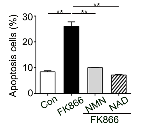
Knowing that the decrease in NAMPT resulted in a decline in NAD+, the researchers injected a NAMPT inhibitor into the healthy mice’s eyes to deplete the NAD+ level. The NAMPT-inhibited-mice exhibited similar symptoms as denervated mice: low NAD+ level and disrupted corneal epithelial balance that causes cell death, also known as apoptosis.
However, replenishing NAD+ levels with the molecule directly or with an NAD+ precursor, nicotinamide mononucleotide (NMN), can rescue the effects of NAMPT depletion. “The percentage of apoptotic cells was significantly lower with the replenishment of NMN or NAD+ and approached equivalence to the normal level,” stated the authors in the study. After injecting NAD+ or NMN into the eye, the level of cell death in NAMPT-inhibitor-treated mice dropped from 25 percent to about 10 percent, which is similar to healthy mice.
NMN Prevents Eye Damage
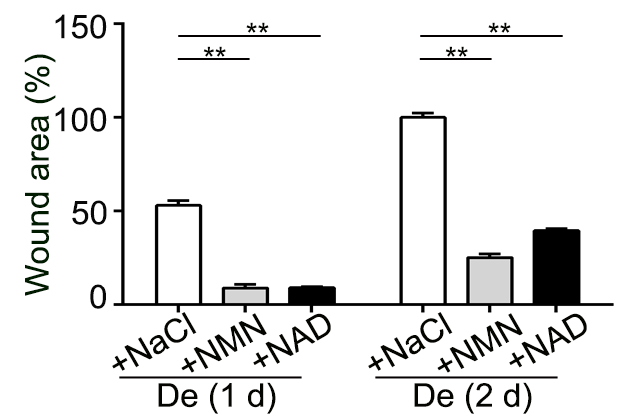
NMN and NAD+ also improved corneal epithelial defects caused by denervation. The replenishment of NMN or NAD+ partially slowed down the loss of the corneal nerve fibers in denervated mice, but it also alleviated epithelial detachment. After two days of NMN or NAD+ treatment, the wound area declined to 25 percent and 40 percent of the original size, respectively.
“Our study demonstrates that corneal denervation impaired the epithelial NAD+ level and caused cell apoptosis and epithelial detachment,” stated the authors. They also suggested that corneal innervations, the process of nerve supply to a body part, play a key role in maintaining the cornea’s stability by regulating NAD+ biosynthesis. “The results suggest that NAD replenishment is a potential treatment to prevent both epithelial shedding and the compromise of corneal function that results from the declines in epithelial innervations.”
 Comments