Research Illuminates Brain Mechanism Involved in Age-Related Motivational Decline and How It Might Be Restored
Elderly individuals often exhibit an age-associated decline in motivation, and researchers from Washington University in St. Louis have unveiled evidence for a brain mechanism underlying it.
Highlights
- Aged mice display reduced behaviors associated with motivation compared to young mice.
- In a brain region crucial for reward and motivation, researchers found lower gene expression of a protein that promotes neuronal survival and growth in aged mice.
- Genetically manipulating young mice to have lower gene expression for this protein recapitulates the reduced motivation observed during aging, suggesting that targeting this protein may be a way to restore motivation with age.
More than 20% of people aged 60 and over have some kind of psychiatric or neurological disorder, with ailments like dementia, depression, and anxiety. Social isolation, a risk factor for such mental health-related problems, is common in older adults as well. Moreover, declining motivation with age can contribute to some adults’ reduced efforts to seek socialization. All the same, aging’s impact on aspects of mental function, like motivation, and the underlying biological mechanisms of motivation in the brain, has remained largely unknown.
Now, as published in Molecular Psychiatry, Shin-Ichiro Imai and colleagues from Washington University in St. Louis showed that aged and socially isolated young mice displayed less motivation-related behavior. Imai and colleagues measured gene expression in a brain region with crucial roles in motivation called the ventral tegmental area (VTA) and found significantly reduced gene expression for a protein called brain-derived neurotrophic factor (Bdnf). Then, genetically manipulating young mice to have lower expression of Bdnf in the VTA significantly reduced their behaviors associated with motivation, recapitulating reduced motivation seen in old mice. These findings suggest that lower Bdnf levels in the VTA play a role in reduced motivation with age and that targeting this protein may serve as a way to restore and maintain motivation during aging.
Background On Bdnf and the Ventral Tegmental Area
Bdnf is a protein that plays a crucial role in promoting the growth and survival of new neurons in the brain, as well as supporting cognitive processes such as attention, memory, and decision-making. Before this study, though, no research had been done connecting reduced Bdnf in the VTA to declining motivation with age.
Moreover, the VTA is a midbrain structure essential for processing rewards (such as pleasurable stimuli), motivation, learning, and cognition through the release of the neurotransmitter dopamine to other brain regions. Furthermore, this brain structure has a central role in influencing behaviors, ranging from basic drive to addiction and even love, by responding to rewards and reward-predictive cues.
Reducing Gene Expression for Bdnf in Young Mice Recapitulates Age-Associated Motivational Decline
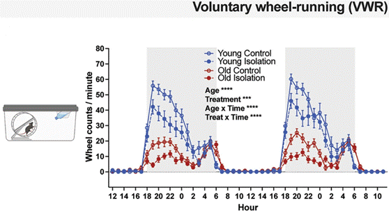
To explore how aging and social isolation influence motivation, Imai and colleagues used young, seven-month-old mice (roughly equivalent to 33-year-old humans) and older, 21-month-old mice (roughly equivalent to 62.5-year-old humans). The mice from these two age groups were exposed to two conditions: one of chronic social isolation (housed alone) and another with group housing conditions (with three to five cage mates). After undergoing these two sets of conditions, the mice were placed on a wheel, where they typically run voluntarily, an innately rewarding behavior even without any other extrinsic reward.
Interestingly, old mice ran much less than young mice during the dark hours of the day, when mice are most active. Moreover, social isolation substantially reduced running time in young and old mice during the dark hours, and this effect was more prevalent in old mice. In that regard, socially isolated old mice ran 40.3% less on average than group-housed old mice, while socially isolated young mice ran 25.7% less on average than group-housed young mice. These results suggest that both aging and social isolation corrode motivation.
Through further experimentation, the researchers sought to parse out the biological mechanisms underlying the observed reduced motivation during aging. Along those lines, extensive scientific research has previously defined the dopamine neurotransmitter system in regulating reward and motivation via a pathway of brain structures that includes the VTA. Two major neuronal populations that produce, store, and release dopamine have been identified in the VTA and another structure called the substantia nigra (SN), both located in the same proximity in the midbrain.
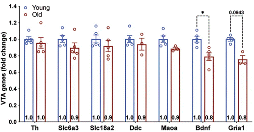
To gain more insight into the mechanism underlying the observed age-related decline in motivation, Imai and colleagues examined the expression levels of genes in these two regions. To do so, they collected tissue samples from both young and old mice and subjected them to a gene expression analysis. The expression analysis revealed significantly reduced gene expression of Bdnf in neurons of the VTA, but not the SN, in old mice.
To confirm that low levels of Bdnf in the VTA play a critical role in reduced motivation with age, the researchers genetically manipulated young mice to have lower Bdnf gene expression in the VTA. With about 30% of VTA neuron Bdnf expression depleted in these genetically-altered mice, the animals were tested for their motivation to seek food. The test they were given required the mice to poke their noses at a location in their cage monitored by an infrared light beam sensor. As such, the mice progressively had to touch their nose to this location more and more times until they ran out of motivation and stopped trying.
Importantly, the genetically-altered mice with lower VTA Bdnf expression exhibited significant reductions in the correct numbers of nose pokes compared to non-genetically manipulated young mice. In that regard, these genetically-manipulated young mice with lower VTA Bdnf exhibited behaviors associated with lower motivation, as seen in typical old mice with reduced motivation.
Increasing NAD+ in the Hypothalamus with eNAMPT Supplementation May Improve Motivation During Aging
In past research, Imai and colleagues had shown that two populations of neurons in a brain region called the hypothalamus (serving as a hub that regulates hormones, sleep, appetite, and other physiological functions) play a vital role in influencing longevity in mice. The researchers are now eager to find whether these hypothalamic neurons also regulate motivation via their connections to Bdnf-expressing neurons in the VTA.
As part of this effort, the research team wants to test whether it may be possible to restore age-associated reduced motivation in mice by supplementing levels of eNAMPT, an enzyme that synthesizes NMN from nicotinamide in the NAD+ biosynthesis pathway. Along those lines, the researchers want to test whether restoring NAD+ levels in the hypothalamus with eNAMPT reverses age-related motivational decline since this brain structure shows lower NAD+ levels with age and since vesicles containing eNAMPT have been shown to extend lifespan in mice. This could mean that eNAMPT supplementation restores hypothalamic function to some degree and that, through the hypothalamus’s connections to the VTA, it could also restore Bdnf in the VTA and, subsequently, motivation.
As for the relevance of this research for people, BDNF levels have been shown to decrease with age in the human brain, and higher BDNF levels have been linked to slower rates of cognitive decline. Moreover, researchers have proposed that lower levels of BDNF may impair synaptic transmission.
As such, it is possible that reduced BDNF in the brain also reduces motivation in people as they age. Accordingly, if supplementing with eNAMPT to increase neuronal NAD+ in the hypothalamus works to restore motivation in aged mice, it may also do so in humans. Currently, the only known method of eNAMPT delivery comes from injections of vesicles containing eNAMPT. These eNAMPT-containing vesicles are not currently available for humans, but if they work to restore motivation in aged mice in future experiments, there may come a time when humans might use them. Of course, vesicles with eNAMPT would need to undergo trials proving their safety first, which could take several years, depending on investments in research on this technology.
 Comments