UC Irvine Scientists Restore Age-Related Vision Loss With a Naturally Occurring Retinal Lipid
Restoring a naturally occurring retinal lipid improves visual function and reduces age-related retinal damage in mice, researchers find.
Highlights
- Supplementation with a retinal fatty acid improved light sensitivity and visual signaling in aged mice.
- Treated mice recovered vision more quickly after exposure to bright light and showed better contrast sensitivity.
- The treatment reduced inflammatory deposits linked to age-related macular degeneration (AMD).
By age 75, nearly one in three adults develops some form of age-related macular degeneration (AMD), a condition that progressively damages central vision. Even in otherwise healthy aging, many people experience declining contrast sensitivity, slower dark adaptation, and reduced visual sharpness long before clinical disease appears. Scientists have increasingly linked these changes to alterations in the retina, particularly shifts in specialized lipids, or fats, that support photoreceptors, the light-sensing cells responsible for vision.
The retina contains unusually high levels of polyunsaturated fatty acids, a class of fats that help maintain the structure and flexibility of photoreceptor membranes. Previous studies have shown that these lipids decline with age and are further depleted in AMD. However, attempts to restore retinal health through dietary omega-3 supplementation have produced inconsistent results, raising questions about whether specific retinal lipids may play a more direct role in vision aging.
In a recent study published in Science Translational Medicine, researchers investigated whether restoring a retinal fatty acid called 24:5n-3 could reverse age-related vision decline in mice. This molecule is produced through the activity of ELOVL2, an enzyme that helps cells produce certain fats and has been strongly linked to aging. The researchers found that aged mice had lower levels of very long-chain polyunsaturated fatty acids in the retina, along with reduced visual performance. Supplementation with 24:5n-3 improved visual function, reduced inflammatory deposits associated with retinal aging, and partially restored gene activity patterns associated with younger retinas.
Aging Retinas Lose Specialized Lipids Required for Vision
The researchers first examined how retinal lipids change with age. In older mice, levels of docosahexaenoic acid (DHA) and very long-chain polyunsaturated fatty acids progressively declined in retinal tissue. These lipids are major structural components of photoreceptor membranes, helping photoreceptors detect light and transmit visual information.
These lipid changes were accompanied by measurable declines in visual performance. Older mice showed reduced contrast sensitivity, weaker retinal responses to light, and slower recovery of vision after exposure to bright light. The animals also accumulated higher levels of inflammatory proteins linked to AMD, including APOE and complement protein C3d beneath the retinal pigment epithelium, a layer of cells that nourishes and supports photoreceptors.
The researchers next studied mice with reduced ELOVL2 activity. These animals developed many of the same retinal lipid abnormalities and visual impairments seen in aging mice, including reduced contrast sensitivity and delayed dark adaptation. The findings suggest that declining ELOVL2 activity may contribute directly to age-related vision loss by disrupting the production of specialized retinal lipids.
Retinal Fatty Acid Supplementation Improves Visual Function
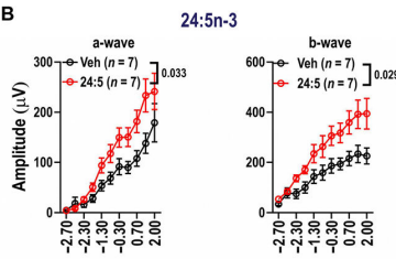
To test whether restoring these lipids could improve retinal function, the researchers injected 24:5n-3 directly into the eyes of aged mice. Five days after treatment, the mice showed improved retinal responses to light under both dim and bright conditions. Treated animals also recovered visual sensitivity more effectively after exposure to intense light, indicating improved function of rod photoreceptors, the cells responsible for vision in low light.
The improvements were specific to aged mice. Young animals did not experience measurable changes after supplementation, suggesting that the treatment primarily corrected deficits associated with aging rather than broadly enhancing visual performance. Other fatty acids tested in the study, including DHA, did not produce the same degree of improvement.
The researchers also measured visually evoked potentials, which assess how visual signals are transmitted from the eye to the brain. Mice treated with 24:5n-3 showed stronger neural responses, suggesting that the improvements extended beyond the retina itself and enhanced overall visual processing. Some benefits persisted for up to four weeks after a single injection.
Supplementation Partially Restores a Younger Retinal Profile
To understand how the treatment affected retinal aging, the researchers analyzed which genes were more or less active after supplementation. Retinas treated with 24:5n-3 showed reduced activity of inflammatory pathways and oxidative stress pathways, which are activated when damaging molecules accumulate inside cells. Genes involved in immune activation and complement signaling, a part of the immune system that helps clear damaged cells and debris, were also less active after treatment.
In parallel, treated retinas accumulated lower levels of APOE and C3d deposits beneath the retinal pigment epithelium. These deposits are commonly associated with retinal aging and AMD development. The researchers found that gene activity patterns in supplemented retinas partially resembled those observed in younger animals, suggesting that the treatment shifted the retina toward a more youthful molecular state.
Human genetic analysis further supported the relevance of the findings. Genetic variants in the ELOVL2 gene were associated with earlier onset of intermediate AMD in two independent human cohorts, suggesting that altered ELOVL2 activity may contribute to retinal aging in humans as well as mice.
Some Aspects of Retinal Aging May Be Reversible
The study suggests that restoring specific retinal lipids may help counter some forms of age-related visual decline. Unlike general omega-3 supplementation, the treatment targeted a lipid directly connected to retinal aging and ELOVL2 activity.
One of the earliest signs of retinal aging is difficulty adapting to changes in light, such as moving from a bright outdoor environment into a dimly lit room or driving at night. Notably, supplementation with 24:5n-3 improved both dark adaptation and retinal responses to light in aged mice. The treatment also enhanced how visual signals were transmitted from the eye to the brain, suggesting benefits that extended beyond the retina itself.
Perhaps the most striking finding was how quickly these improvements occurred. Within days of treatment, aged mice showed measurable gains in visual function, and some benefits persisted for up to four weeks after a single injection. Although the work was performed in mice and used intravitreal injections, which deliver treatment directly into the eye, the results suggest that some age-related changes in vision may not be inevitable consequences of aging and could potentially be reversed by restoring key molecules lost over time.
Model: Aged wild-type male C57BL/6J mice
Dosage: Mice received short- and long-term intravitreal (inside-the-eye) injections of 24:5n-3. The study does not specify the injection concentration or dose in the primary Methods section.
 Comments